
Abstract
Biotin is a universal molecule that is essential for the metabolism of all life forms. Biotin acts as a coenzyme for carboxylases, playing a crucial role in various metabolic pathways including gluconeogenesis, amino acid catabolism, and fatty acid synthesis. Beyond these functions, biotin also influences gene expression regulation.
The essential role of biotin for life extends back to the beginning of life, and even prebiotic metabolic organizations. This molecule has remained largely unchanged throughout the history of life, indicating its evolutionary conservation. The diversity and conservation of biotin-dependent enzymes highlight their significance in metabolic flexibility and adaptation.
Keywords: Biological Evolution, Biotin, Carboxylases, Coenzymes, “Metabolism, Inborn Errors”, Vitamins
INTRODUCTION
Biochemical-Clinical Characteristics of Biotin
Chemical Properties of Biotin
Biotin is a water-soluble vitamin and a universal molecule required for the metabolism of all living organisms. It is also referred to as vitamin H, vitamin B7, or coenzyme R. Biotin remains stable at room temperature and is not broken down by cooking. The term “biotin” comes from the Ancient Greek words “biotos” (meaning “life”) and “-in” (derived from a common suffix used in chemistry).
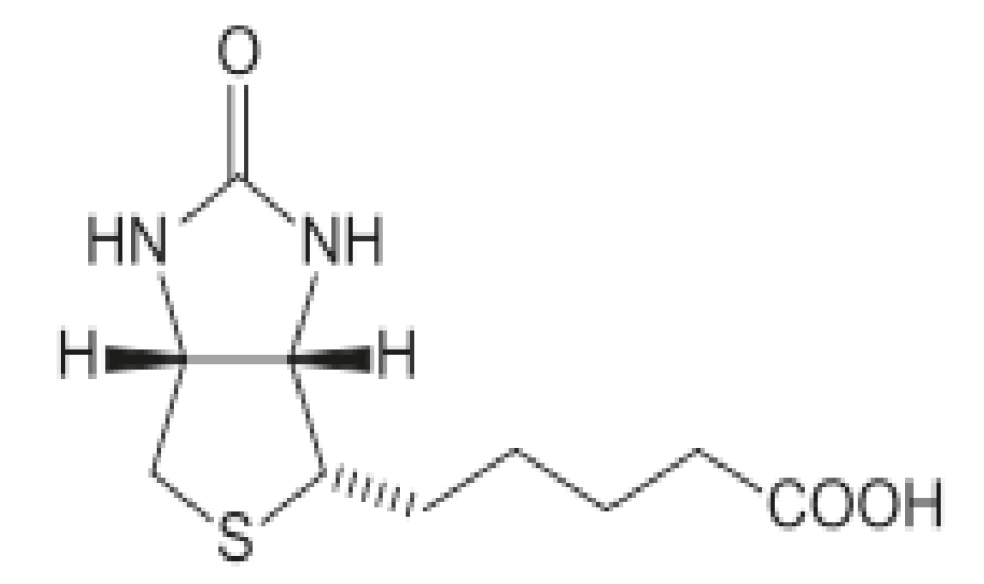
Chemically, biotin is a prosthetic group consisting of a valerate side chain attached to a bicyclic ring made up of a ureido and a thiophane ring (). The molecule is a combination of a tetrahydroimidazolone ring and a tetrahydrothiophane ring containing an organosulfur group bearing a valeric acid substituent. Biotin is covalently linked to the protein through a lysine residue.
History of the Discovery of Biotin
The discovery of the biotin molecule and research on its properties are detailed in .
Table 1. History of the discovery of biotin
1898 | Discovery of toxicity of raw egg whites (Steinitz) |
|---|---|
1916 | A diet rich in raw egg whites causes toxic symptoms in dogs, cats, rabbits and humans (WG Bateman) |
1927 | Egg white damage. Neurotoxicity, alopecia, dermatitis in rats fed only egg whites (M. Boas and H. Parsons) |
1936 | Isolation of crystallized biotin. Named yeast growth factor (Fritz Kögl and Benno Tönnis) |
1939 | Discovery of factor responsible for egg white damage. Named vitamin H (P. Gygory, representing Haar und Haut) Named coenzyme R in experiments with yeast and Rhizobium trifolii (West Wilson) |
1940 | The name biotin was used for the first time. It was discovered that the damage was due to the binding of biotin to avidin (Gygory) |
1950 | Discovery that biotin plays a role in carboxyl transfer |
1968 | The synthesis pathways for biotin in bacteria (Escherichia coli) were discovered |
Biotin Physiology and Metabolism
Unlike bacteria and plants, higher mammals cannot synthesize biotin endogenously, so they must obtain it from the diet. Biotin taken from foods in the diet is found in free form or bound to proteins. Absorption of biotin from the diet is quite effective, even when taken in large amounts. Therefore, its deficiency is quite rare. Biotin taken with food is optimally utilized through a cyclical system known as the biotin cycle. In this cycle, biotin is used in metabolic pathways and then returned to the circulation to be reused (). When biotin is consumed with food, it is broken down into biocytin (biotinyl-ε-lysine) with the assistance of proteolytic enzymes in the digestive tract. In pancreatic and intestinal secretions, biotinidase (BTD) located on the brush borders of the apical surface of enterocytes releases biotin.
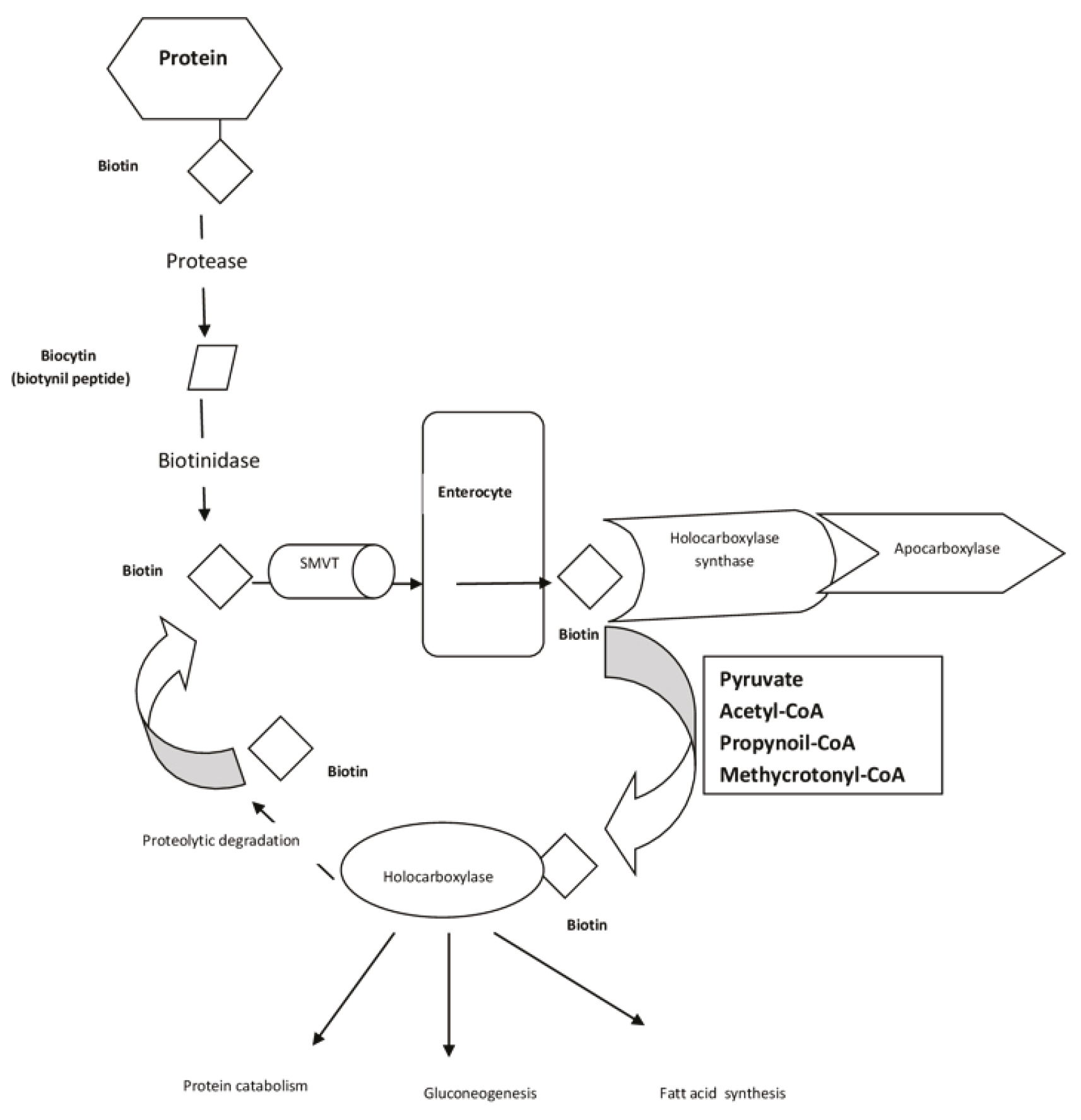
SMVT: Sodium-dependent multivitamin transporter
With the assistance of the sodium-dependent multivitamin transporter (SMVT) enzyme, free biotin is absorbed from the apical surface of enterocytes as an electroneutral substance. It then enters circulation as an electrogenic solution from the basolateral surface, independently of sodium. Because SMVT is highly sensitive to pantothenic acid and lipoic acid, intake of multivitamins may competitively affect biotin uptake. However, an important way animals acquire biotin is through synthesis by bacteria found in the intestinal microbiota. Unfortunately, the microbial synthesis in the intestines is not sufficient to meet metabolic needs due to the lack of necessary transport proteins. The normal gut flora and normal intestinal production of biotin in an untreated individual with profound BTD deficiency mean that the affected individual will develop secondary biotin deficiency and multiple carboxylase deficiency. If the biotin produced in the flora were adequate, these individuals would likely not become symptomatic. This fact confirms that biotin synthesized by bacteria in the human intestines is insufficient to meet the requirements in these cases.
Biotin that enters the circulation is then transported to the liver and other peripheral tissue cells with the assistance of SMVT. Reabsorption of biotin from the kidney glomeruli is also facilitated by SMVT. Biotin binds to holocarboxylases, which activate carboxylases in the cell and enable their function. Biotin is subsequently catabolized through two degradation pathways: biotin beta, involving the breakdown of the valeric acid side chain and sulfur oxidation, resulting in the formation of bisnorbiotin, biotin sulfoxide, and other metabolites, which are then excreted in the urine. The remaining biotin is reabsorbed from the kidney glomeruli via SMVT and re-enters the biotin cycle.
Biotin’s Biological Functions
Biotin serves two roles: as a coenzyme and as a non-coenzyme. Its primary function is to act as a carboxyl carrier in carboxylation reactions. Biotin’s main role is to supply CO2 to carboxylase enzymes, which are found universally in all three branches of life. Carboxylase, decarboxylase, and transcarboxylase enzymes, crucial in various metabolic processes such as branched-chain amino acid catabolism, gluconeogenesis, and fatty acid synthesis, require biotin for their activities. Mammals have four biotin-dependent carboxylases: acetyl-CoA carboxylase, pyruvate carboxylase, propionyl-CoA carboxylase, and b-methylcrotonyl-CoA carboxylase. Biotin serves as the universal coenzyme for these enzymes. Biotin serves as a coenzyme responsible for transferring bicarbonate to acetyl-CoA, which is then converted to malonyl-CoA for fatty acid synthesis. Pyruvate carboxylase plays a role in gluconeogenesis, while b-methylcrotonyl-CoA carboxylase catalyzes a step in leucine metabolism. Additionally, propionyl-CoA carboxylase catalyzes a step in gluconeogenesis.
Evidence is accumulating on the important role of biotin as a non-coenzyme. It also plays a role in epigenetic mechanisms, such as biotinylation of histone proteins, stability of nuclear chromatin, and gene expression. Additionally, biotin is effective in cell signaling. More than 2000 biotin-dependent genes have been identified, and there is evidence that bisnorbiotin and biotin catabolites affect gene expression. Research has shown that biotin causes a six-fold downregulation of phosphoenol pyruvate carboxykinase expression.
Based on these findings, studies have been conducted on the treatment of diabetic rats with biotin. It has been reported that biotin affects ornithine transcarbamylase activity, and its deficiency, biotin deficiency, is accompanied by hyperammonemia.
Biotin in the Diet
Biotin is widely found in foods, with absorption levels varying. Offal contains high levels of biotin, while grains contain less. Daily intake recommendations differ based on age, with 5 µg/day recommended for newborns and 35-70 µg/day for adults. No toxicity from nutritional deficiencies or excessive biotin intake has been reported.
Certain conditions can impact daily biotin requirements. These include pregnancy, long-term total parenteral nutrition, severe and prolonged malnutrition, high egg white consumption, chronic alcohol use, use of antiepileptics, use of multiple vitamins containing lipoic acid, gastrectomy, and achlorhydria.
There are many causes of biotin deficiency. It can occur in inborn errors of metabolism, which are rare, including BTD deficiency, carboxylase deficiencies, holocarboxylase synthetase deficiencies, and carrier protein deficiencies. BTD deficiency is a common metabolic disease globally, leading to a decrease in enzyme activity, which results in multiple carboxylase deficiency, resulting in inadequate biotin recycling and utilization.
Clinical Finding in Biotin Deficiency
The most severe symptoms, including hypotonia, convulsions, developmental delays, ataxia, coma, and hearing loss, can manifest in biotin deficiency. Dermatological symptoms may include alopecia, rashes, periorificial dermatitis, conjunctivitis, thin and brittle hair, and brittle nails. Immunodeficiency symptoms may involve susceptibility to candida infections, low lymphocyte subgroups, and impaired antibody synthesis.
Biotin deficiency may also be linked to lipid metabolism disorders, resulting in high levels of single-chain fatty acids. Additionally, there have been reports suggesting that biotin deficiency could be teratogenic. During pregnancy, biotin deficiency may lead to conditions such as cleft palate-lip, micrognathia, and micromelia.
Laboratory Findings in Biotin Deficiency
Biotin deficiency can result in keto-lactic acidosis, hyperammonemia, and organic aciduria.
Diagnosis in Biotin Deficiency
Serum and urine biotin levels are not reliable for diagnosis. Urinary 3-hydroxy isovaleric acid excretion can be helpful for diagnosing metabolic disorders. It is important to note that biotin can interfere with laboratory tests, particularly those using biotin-streptavidin technology for measuring thyroid hormones and vitamin D levels.,
Use of Biotin in Diseases
Biotin may be used for experimental purposes or in certain proven cases. Some studies suggest that high doses, such as 300 µg/day, may impact the progression of multiple sclerosis. However, many subsequent studies have shown that biotin has no effect on multiple sclerosis., In animal studies, biotin has been shown to increase insulin secretion in diabetic mice. It is also recommended for improving hoof health in animals and hair health and nail health in humans.,
Future Perspectives for Biotin-Related Research
Research on fundamental biomolecules such as “coenzymes” and especially “biotin” will lead to promising developments in the future, both in terms of evolutionary biology and in scientific/applied fields.
Evolutionary Importance
Evolutionary conservation of cofactors: Coenzymes [nicotinamide (NAD)⁺, flavin adenine dinucleotide (FAD), coenzyme A, biotin, etc.] have been used since the earliest stages of life. For example, biotin is essential for the functioning of carboxylase enzymes. The preservation of such molecules from common ancestors to the present demonstrates that they are among the “universal constants” of biochemical evolution.
Evolution of metabolic pathways: Coenzymes like biotin are involved in key points in carbon metabolism (e.g., gluconeogenesis, fatty acid biosynthesis). Metabolic innovations that increase energy efficiency throughout evolution have often depended on these coenzymes.
Future perspective: New environmental pressures (climate change, artificial feeding environments, space life) will test the metabolic flexibility of organisms. The bioavailability and synthesis of essential coenzymes like biotin may be critical to the adaptive success of species in the future.
Importance in Scientific and Applied Fields
Biotechnology: Biotin is a unique tool for labeling biomolecules through the “avidin-biotin” system. This system may be more widely used in nanotechnological biosensors, drug delivery systems, and sensitive diagnostic kits in the future. In metabolic engineering, biotin-dependent enzymes can be manipulated to construct artificial biosynthetic pathways.
Medicine and nutrition: The role of biotin deficiency in the nervous system, metabolism, and epigenetics is increasingly being investigated. Its links to neurodegenerative diseases, diabetes, and obesity may become particularly important.
The role of coenzymes (such as NAD⁺ and sirtuins) in epigenetic regulation has been demonstrated. Biotin is also known to affect gene expression through histone biotinylation. This could be a target for future epigenetic therapies.
Space biology: Metabolism will be subjected to different stresses during long-term space travel. The stability, synthesis, and recycling of coenzymes may become critical in life support systems.
Future Scenarios
Synthetic biology: Biotin and similar coenzymes can be used as “modular metabolism tools” in artificial or genetically engineered organisms.
Evolutionary engineering: During laboratory evolution of organisms, selection can be applied to coenzyme metabolism to impart new biochemical properties.
New treatments: Biotin and other coenzymes may be central to both drug development and personalized nutrition approaches in the future.
In the future, biotin may be used as a mitochondrial function supporter or neuroprotective agent.
Plant biotechnology: Biotin is critical for photosynthesis and carbon fixation. Genetic engineering can enhance biotin metabolism to develop drought-resistant, productive crops.
Livestock: Biotin deficiency impairs coat/skin health. In the future, highly bioavailable feed additives may improve both animal welfare and productivity.
Functional foods: Biotin-fortified foods (especially in older adults and metabolic risk groups) may become widespread.
To summarize, coenzymes have a remarkably stable evolutionary role as the “common chemical language” of life. Biotin, in particular, appears poised to remain a strategic molecule in both the evolution of metabolic networks and in biotechnological and medical applications in the near, medium, and long-term.
Possible Evolutionary Insights on the Place of Its in the Origin of Life and the Metabolic Organization
Biotin is a universal coenzyme with an evolutionary origin that dates back to the emergence of life and even before. The role of coenzymes and cofactors in the origin of life which act as catalysts in biochemical reactions, has been largely overlooked until now. Considering the initial conditions of the Earth, the role of coenzymes in the evolution of inorganic molecules into organic molecules, specifically in abiogenesis, is of critical importance. Coenzymes have played a significant role in the evolution of metabolic pathways by extending from proto-metabolism to prebiotic metabolism, and from there to biotic metabolism. The suggestion that amino acid biosynthesis cannot occur without coenzymes and cofactors. Coenzymes emerged on Earth before the evolution of the Last Universal Common Ancestor (LUCA). To gain a comprehensive understanding of the evolution of biotin as a coenzyme, it is necessary to consider the geological evolution of the Earth, the emergence of metabolism, and the emergence of the cell. The emergence of coenzymes, including biotin, is interconnected with the evolution of amino acids, proteins, nucleic acids, metabolism, and the cell, indicating a coordinated evolution.
Evolutionary Insights on Origin of Life and the Metabolic Organization
Geological Chronology
The observable universe dates back approximately 13.7 billion years. The Earth began to form approximately 4.6 billion years ago with the accumulation of masses that accreted from the solar nebula.
At its beginning, the Earth was quite different from today. There was no atmosphere or water yet. Intense meteor bombardment and tectonic activity were common. Active volcanic activities, involving magma and lava, produced the electrical energy required for life to emerge through non-enzymatic means. It is suggested that water was carried to Earth by continuous asteroidal bombardment. During this time, Earth was a very hot planet, so the water was in the form of water vapor. Over time, the atmosphere and oceans began to form as a result of changes in the composition of gases. Initially, the atmosphere and oceans were a toxic environment rich in H2, N, C, Fe, S, NH3, and CH4. The atmosphere was primarily rich in N, but over time, O2 levels increased while N levels decreased. The cycles of N, O2, and C formed the biosphere.,
The continents had not yet formed, and the land mass was a single piece called Pangea. The Moon was formed by the merging of pieces that broke off from Earth when a planet called Theia collided with Earth about 50 million years after Earth’s formation.,
Chronology of Life
The first proto-cells on Earth evolved approximately 1 billion years after the formation of the Earth, around 3.5-3.6 billion years ago. According to the widely accepted view, the first cells emerged at the base of hydrothermal vents on the ocean floor. There are debates surrounding the definition of life. One of the most well-known definitions of life is as follows: Life is any self-sustaining chemical system capable of Darwinian evolution. Life involves the possession of dynamic information about organized relationships between material entities ().
Table 2. Evolution of metabolism and life based on Earth geological periods
Geological evolution | Evolution of metabolism | Evolution of life |
|---|---|---|
- | Hadean period (4.6-3.8 billion years ago) | - |
Intense meteor bombardment and tectonic activity, lava, magmas - Formation of reducing atmosphere • H2, CO2, CH3, SO2, NH3 - Formation of hydrothermal vents in oceans • C, CH4, S, NO, Fe | Prebiotic-protobiotic-abiotic metabolism C, H, N, S, Fe, cycles (O2 is very low) Non-enzymatic, anaerobic metabolism Inorganic chemical reactions Primordial soup Chemical reaction rate is very slow Energy source (sun, lightning, ultraviolet, volcanic lava) | Inorganic molecules Coenzyme/cofactors (biotin, RNA, DNA, Ribozyme Coacervates Chemical synthesis of amino acids - RNA world hypothesis/gene first Metabolism hypothesis/metabolism first Coenzyme hypothesis |
- | Archaean period 3.8-2.5 billion years ago | - |
In the atmosphere • CH4, NH3, toxic gases, H2, CO2, CH3, SO2 - In the oceans • C, Fe,S, NO | Cellular-biotic metabolism Anaerobic photosynthesis Chemoautotroph-photoautotroph Autotroph metabolism (capable of creating its energy from inorganic molecules) Heteretroph metabolism (capable of synthesizing its energy from organic structures) Metabolic synthesis of organic molecules aa Emergence of complex metabolic pathways Very fast (discovery of enzymes) Energy source (electrochemical forces-enzyme) | Protocells - evolution of cells Cell membrane Enzymes Evolution of archaea and bacteria Chemical autotrophs (in the ocean) Photosynthetic cyanobacteria (O2 in the atmosphere) Evolution of the cell Last Universal Common Ancestor 2 |
- | Proterozoic era 2.5 billion-541 million years ago | - |
Great oxygenation event - Increase in O2 in the atmosphere, decrease in N - Decrease in Fe, S, N in the ocean | Anaerobic photosynthesis Chemoautotroph Photoautotroph Nitrogenase, formation of nitrogen cycles Endosymbiosis Interconnection of complex metabolic pathways Krebs cycle Urea cycle | Evolution of eukaryotes Evolution of multicellular life Colonial life Early animals (hydras, sponges) Bilateral animals Chordalians Vertebrates |
- | Paleozoic era 541-252 million years ago | - |
Cambian Ordovician Silurian Devonian Carboniferous Permian | - | Cambrian explosion Increased biodiversity in vertebrates Plants emerging onto land Tetrapods emerging onto land Evolution of the amniotic egg |
- | Mesozoic era 252-66 million years ago | - |
Triassic Jurassic Cretease | - | The emergence of the first mammals dinosaurs The emergence of flowering plants |
- | Cenozoic era 66 million years ago - present | - |
Paleogene Neogene Quaternary | - | Evolution of primates Hominins Homonoids Homo sapiens |
Initially, cells emerged as a result of the coordinated development of life characteristics, including compartmentalization (membrane), metabolism, and self-replication (genetics), during millions of years of evolution. Life is an intermediate stage that has gained the ability to renew itself; this is brought about by the chemical kinetic selection resulting from reduction-oxidation (redox) reactions during the global chemical cycle between the most common elements C, H, O, N, S, and Fe found in nature after the formation of the world. Life emerged at each level through the natural evolution of compounds that underwent chemical selection due to the potential for further reproduction, with each simple reaction or event cycle forming a product that activates itself, leading to increasingly complex structures each time.
The question of how life emerged from inorganic molecules under the initial conditions of the Earth, remains a subject of lively scientific interest. There are several significant hypotheses on the subject, including the RNA hypothesis, metabolism hypothesis, and coenzyme hypothesis.
RNA/Gene First Hypothesis
The RNA/Gene First Hypothesis suggests that self-replicating components emerged before metabolism. In the 1960s, Cairn Smith proposed the “clay life model,” which posited that clay may have been the first genetic material capable of replicating itself through crystallization. In this model, clay-based life materials could have utilized organic materials in the environment to create genes.
Another popular model within the gene first approach is the RNA world hypothesis, first introduced by Walter Gilbert. This hypothesis suggests that RNA and DNA appeared before proteins. Initially, RNA molecules had structures that allowed for self-replication, information storage, and enzymatic properties. These RNA structures with enzyme-like properties are known as ribozymes. Evolutionarily, RNA is considered the ancestral molecule of DNA.
Ribose, a sugar found in RNA, is more unstable compared to deoxyribose, a sugar found in DNA that emerged later and is more stable. The first evolved precursor RNA molecule was tRNA. Coenzymes and cofactors, such as biotin, were more commonly associated with RNA rather than proteins.
Metabolism-First Hypothesis
According to the metabolism first hypothesis, the Earth’s conditions initially hosted a series of chemical reactions that created molecules that would eventually give rise to life. The hypothesis was first proposed in 1924 by Russian scientist Alexander I. Oparin, known as the “coacervate theory”. This theory suggested that iron carbides chemically reacted with water vapor, leading to the formation of hydrocarbons, alcohols, aldehydes, and other organic chemicals. These compounds then combined with NH3 to form amides, amines, and other NH3 compounds, creating colloidal structures known as coacervates, which are protein precursors.
Independent of Oparin, the renowned British scientist JBS Haldane named the environment, where he proposed life began on Earth, the “primordial soup” in 1929. Inspired by these theories, scientists Stanley Miller and Harold Urey demonstrated in 1953 that amino acids could be synthesized from biomolecules by simulating the early Earth conditions, sparking further interest in the hypothesis. The experiment replicated the electrically charged environment with spark discharges by heating a glass bottle containing a mixture of water, methane, hydrogen, and ammonia, and continuously stimulating it with electric sparks.
In the 1980s, Gunter Wachtershauser reinforced the credibility of the metabolism hypothesis by proposing the “surface metabolism theory. Wachtershauser demonstrated that protein precursor structures could self-replicate through autotrophic mechanisms on electrically charged iron sulfide mineral surfaces, such as pyrite, without the need for a cell membrane. This theory suggested that porous rock surfaces in hydrothermal vents in the ocean functioned as protocells without a membrane.
Today, the theory of abiogenesis, the formation of amino acids and proteins from inorganic molecules under Earth’s conditions, has been extensively detailed. It is now widely accepted that life did not emerge all at once through a transcendent abstract force but evolved through biochemical processes over billions of years, transforming chemistry into biology.
Coenzyme Hypothesis
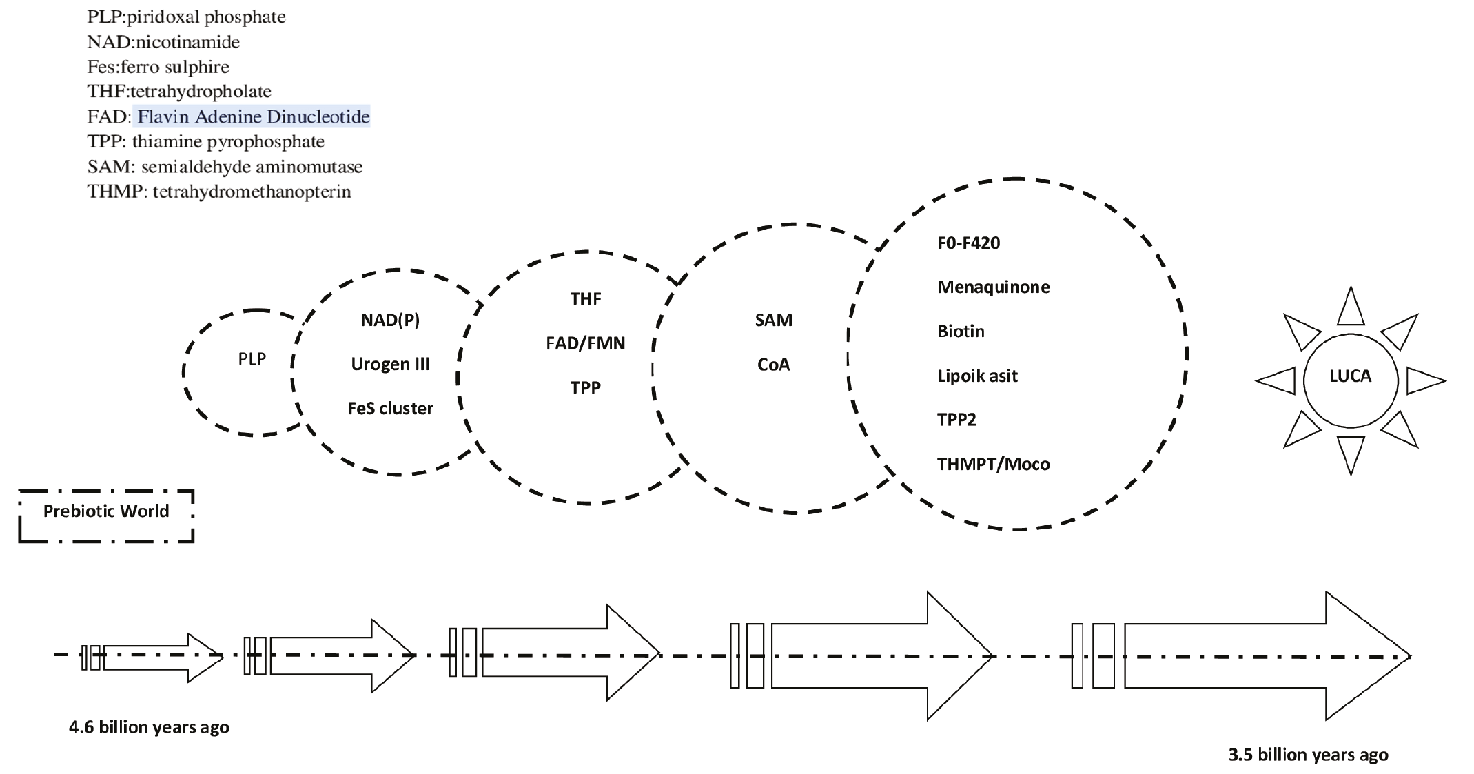
The coenzyme hypothesis suggests that life originated with the help of molecules like biotin, pyridoxal phosphate, thiamine pyrophosphate, adenosine triphosphate (ATP), AGP, siroheme, ferredoxin, ferro sulphire, NAD, FAD, pterin etc. (). These molecules are similar to coenzymes and are also simple, catalytically active, self-replicating, and capable of facilitating chemical reactions. They were believed to have settled on the surface of oil droplets in water, altering the surface properties, and allowing for self-replication. The evolution of early systems relied on the collaboration of numerous self-replicating molecules and the development of self-organization among them. Without these molecules, the emergence of organic molecules such as coacervates, amino acids, and proteins would not have been possible.
The Evolution of Metabolism
Metabolism is a network of processes that repeat and sustain themselves by regulating the flow of matter and energy that gives life to molecular structures. Current metabolic pathways are not simply the sum of their parts. Parts cannot exist independently of the process that creates the whole, as the whole and the parts are the product of a dialectical process of interdependence. Like all other biological phenomena, the current location and state of metabolism depend not only on its current formation, but also on a past that gives rise to different possibilities for the present and future interactions of its parts. As the famous evolutionist Theodosius Dobzhansky said, “Without the light of evolution, nothing in biology has meaning.” No matter how complex metabolic pathways may seem, when the uninterrupted chain where each is connected to the previous is followed, it will be seen that self-organization is at work. The driving force required for the evolution of complex metabolic pathways can be found in the second law of thermodynamics. All systems far from equilibrium are necessarily pushed towards more complex structures as long as there is a flow of energy and substrate (dynamic-kinetic stability), formulated by Nobel Prize-winning scientist Ilya Prigogine, who was inspired by this law.
The origin of metabolism can be traced in two stages: the prebiotic period, when cells had not yet emerged, and the post-cellular biotic period, ().
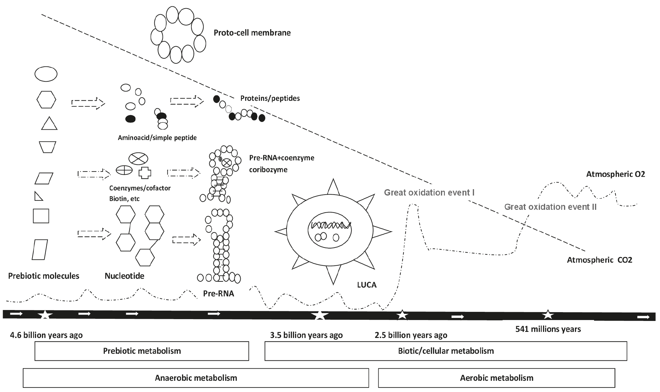
LUCA: Last universal common ancestor
Prebiotic, Proto-Biotic, Abiotic Metabolism (4.5-3.8 Billion Years Ago)
It was a phase in which organic molecules, derived from inorganic molecules, formed the basis of life, leading to the formation of protocells. This period, known as the Hadean period, was characterized by a reducing atmosphere that eventually gave way to cycles involving carbon, nitrogen, sulfur, iron, and hydrogen. With oxygen not yet dominating the atmosphere, anaerobic metabolism was prevalent. Energy for chemical reactions was derived from sources such as the Sun’s ultraviolet rays, lightning, and volcanic lava. Organic molecules were primarily formed at the hydrothermal vents at the ocean floor. Coenzymes, such as amino acids, coacervates, and biotin, were among the substances that emerged during this period, along with precursor molecules for RNA and DNA, whereas NH3 is not a coenzyme.
Cellular-Biotic Metabolism (3.8-2.5 Billion Years Ago)
This is the stage when the first cells, LUCA, emerged. Archaea and bacteria evolved. These are cells with autotrophic metabolism that perform anaerobic photosynthesis using Fe and S. Later, cells with heterotrophic metabolism evolved. Thus, cells that can synthesize their energy from organic structures, that is, cells with cellular biotic metabolism, emerged. Complex metabolic pathways were interconnected with each other. During this period, changes in the C, H, O, N, S, P ratios that emerged in the atmosphere and ocean strengthened the evolutionary selection pressures that led to the revelation of cellular heterotrophic metabolism in life forms. Among the pressures that shaped metabolism, the ability to use newly emerged resources (anabolism) and the avoidance of toxic wastes and protection from them were decisive. Metabolism is shaped by the sum of the responses that organisms give to the environment. Therefore, what constitutes metabolism today also requires an understanding of the evolutionary geology that created it., The environmental pressures that led to the evolution of cellular metabolism, that is, the transition from autotrophic to heterotrophic metabolism, are as follows:
• Depletion of energy and nutrient source chemicals in the primordial soup.
• Change in the nitrogen cycle (nitrogen crisis). Decrease in reduced nitrogen in the ocean.
• Increase in O2 in the atmosphere (Great Oxigenation Event, GOE). As a result of the pressure to protect from the toxic effects of oxygen and increase the ability to use oxygen, aerobic photosynthetic cells were selected.
• C change in the atmosphere. The need for fixation of atmospheric carbon into organic structures. Here, the evolutionary use of carboxylases and their coenzyme biotin came into play. As a result, the evolution of complex metabolic pathways freed cells from chemical dependence on essential prebiotic molecules.
Evolution of LUCA
LUCA is a term used to describe the universal cell community that is considered the ancestor of the three main branches in the tree of life: archaea, bacteria, and eukaryotes. It is believed to have evolved approximately between 3.6 and 2.5 billion years ago. Common metabolic pathways found in all cells are thought to have originated from LUCA. Rather than being a single cell, LUCA is considered a community of cells where universally common genetic elements that can be expressed and copied are present. It is believed to have had a genome consisting of 1,000-1,500 ancestral genes. Eukaryotes are thought to have evolved around 2.5 billion years ago as a result of cells with different characteristics combining through endosymbiosis after the increase in atmospheric O2 (GOE) and the subsequent increase in biodiversity.
Possible Evolutionary Insights on the Role of Biotin as a Coenzyme in the Origin of Life and Metabolic Organization
The Evolutionary Importance of Coenzymes
Coenzymes are organic molecules that play a crucial role in energy conservation by reducing the activation energy needed for biochemical transformations. As a result, they are universal molecules that have been essential in the origin of life and continue to be vital in cellular functions. Coenzymes hold significant evolutionary importance and are believed to be remnants of the prebiotic RNA world, sharing structural similarities with RNA. They are present in all three parallel lineages of molecular and metabolic evolution and can form partnerships with nucleic acids, enzymes, and proteins.
Remarkably, coenzymes have undergone minimal structural changes since the LUCA and have been well-preserved throughout evolution. This preservation allows them to serve as control points in molecular evolution, playing crucial roles in proto-metabolism and current metabolism. Despite their importance, coenzymes are relatively few in number, which limits the scope of molecular evolution.
Genes involved in coenzyme biosynthesis, such as biotin and vitamin C, have been gained and lost in various life forms over time. The evolutionary origins of coenzymes date back even before LUCA, highlighting their critical role in the development and maintenance of life processes.
The Evolutionary Importance of Biotin
When considering the role of biotin from biochemical and genetic perspectives, this consideration sheds light on the origin and diversity of life. Biotin is a universal molecule that plays an essential role in the evolution of life preserved in the three main branches of life alongside lipoic acid. Biotin has been crucial in the origin of life, serving as a central player in the fixation of biospheric carbon into organic structures and as the key molecule for CO2 transfer.
Biotinylated enzymes, known as carboxylases, are vital in metabolism across all three branches of life and are dependent on biotin. Evolution is intertwined with biotin, a key coenzyme in basic and intermediate metabolic pathways. Many pathways related to carbon metabolism require biotin, although CO2 transfer can occur without it. Biotin, along with lipoic acid, evolved relatively late, and both share structural homology.
Before biotin, RuBisCo (Ribulose-1,5-bisphosphate carboxylase/oxygenase) played a role in fixing CO2 from the atmosphere. It is one of the most abundant enzymes on Earth. A mutual and symbiotic relationship has developed between biotin-synthesizing bacteria and plants, highlighting the importance of biotin for metabolic flexibility in most life forms. Species have successfully adapted to this diversity. In addition to its evolutionary roles, biotin also plays a role in gene expression regulation.
Bacteria, plants, and some fungi are capable of synthesizing biotin. A biotin-binding protein binds biotin to carboxylases. This enzyme acts as both a ligase and a transcription factor, indicating an early evolutionary mechanism for regulating biotin metabolism. Mammals, on the other hand, cannot synthesize biotin as they have lost the biosynthesis gene required for it. The biosynthesis of biotin is a costly process, requiring about 19 ATP molecules and at least 6 enzymes to produce just 1 biotin molecule. Therefore, mammals rely on obtaining biotin from their diet and/or intestinal microbiota. This highlights the importance of biotin in symbiotic relationships during evolutionary processes. Acquiring biotin from the environment is more energy-efficient than synthesizing it. The loss of the biosynthesis gene is a well-documented evolutionary phenomenon, driven by energy costs. Genes involved in biosynthesis are often lost when alternative, more cost-effective methods of obtaining essential molecules are available.
There are numerous examples of this evolutionary phenomenon, such as the loss of essential amino acid synthesis genes in eukaryotes with symbiotic life 850-650 million years ago, the loss of the vitamin C synthesis gene after the consumption of plants, 60 million years ago, and the loss of the uricase gene, whereby the pathway evolved for converting fructose from fruits, into fatty acids for energy gain 25 million years ago.- Additionally, the loss of 85 genes that facilitated adaptation to aquatic life was observed during the transition from aquatic to terrestrial. The endogenous biosynthesis of vitamin D became less significant with reduced sun exposure following the exodus from Africa 70,000 years ago, making vitamin D essential and it must be obtained externally.
The BTD enzyme is found in everything from prokaryotes to fungi, from arthropods to mammals. The evolution of BTD dates back to the beginning of life. BTD genes in various species have regions containing specific amino acids that are evolutionarily conserved, and these regions show high homology between species.
Evolution of Biospheric Carbon Fixation
The fixation of inorganic carbon compounds, such as CO2, into more reduced organic forms is one of the most fundamental processes of life. This fixation is a prerequisite for life and serves as the starting point of biological evolution, placing the biosphere within geochemistry. Many prokaryotes and all plants have a dominant mechanism, the Calvin-Benson cycle, by which they fix CO2 into biomass. The diversity in carbon fixation forms the molecular basis of many deep branches on the tree of life, leading to metabolic diversity that traces back to the earliest cells. The evolution and productivity of carbon fixation pathways have also been influenced by changes in oxygen and carbon concentrations throughout geological time.
Acetyl CoA is a critical molecule that plays role in the fixation of atmospheric carbon into organic structures. It serves as the first step in most metabolic pathways, including gluconeogenesis, and acts as the common currency of metabolic processes. Acetyl CoA is the primary building block of carbon-based life forms, marking the beginning of the carbon-based backbone of life. Acetonogens and methanogens utilize acetyl CoA for H2-dependent carbon and energy metabolism, forming acetate through the autotrophic fixation of CO2.
Evolution of Biotin-Dependent Carboxylases
Carboxylases are present in all three main branches of life and can be traced back to the LUCA. They exhibit high homology across different species. Biotin-dependent carboxylases have been known to exist since the early stages of life, particularly for pyruvate carboxylation and acetyl CoA carboxylation, highlighting the crucial role of biotin in metabolic processes. Despite the diversity of various life forms, their fundamental functions have remained consistent. They are essential components of biological evolution and metabolism, playing a key role in incorporating atmospheric carbon into organic structures during the evolutionary process. Due to their involvement in CO2 transport, they are vital in all intermediate stages of metabolism.
The last common ancestor of archaea possessed two biotin-dependent carboxylases, while the last common ancestor of bacteria had three. Eukaryotes likely acquired biotin-dependent carboxylases through symbiotic relationships, such as endosymbioses with mitochondria and plastids, as well as from other unknown bacterial sources. While some bacteria and archaea have evolved the ability to synthesize biotin, organisms like humans must obtain it through their diet, underscoring the evolutionary significance of symbiotic interactions.
It has been proposed that the carboxylase family evolved from small, single-function precursors to generate multifunctional polypeptides through duplication, amplification, and recombination events. Prior to the emergence of carboxylases, RuBisCo played a key role in carbon fixation.
CONCLUSION
Biotin is a universal molecule that is essential for the metabolism of all life forms. The evolution of biotin provides insight into the origin of life, reaching back to a time before life itself. Biotin acts as a coenzyme for carboxylases, playing a crucial role in various metabolic pathways including gluconeogenesis, amino acid catabolism, and fatty acid synthesis. Beyond these functions, biotin also influences gene expression regulation.
The evolution of biotin dates back to the beginnings of life and even to prebiotic metabolic processes. This molecule has remained largely unchanged throughout the history of life, indicating its evolutionary conservation. The diversity and conservation of biotin-dependent enzymes highlight its significance in metabolic flexibility and adaptation.
For organisms like humans that cannot produce biotin, reliance on intestinal microbiota and dietary sources has emerged as an evolutionary adaptation. Although it is synthesized by bacteria in the gut, mammals have lost the ability to produce biotin internally due to the high cost of biotin biosynthesis and require external sources.
Footnotes
Authorship Contributions
Surgical and Medical Practices: M.K., Y.K., Concept: M.K., Y.K., Design: M.K., Y.K., Data Collection or Processing: M.K., Y.K., Analysis or Interpretation: M.K., Literature Search: M.K., Y.K., Writing: M.K., Y.K.
Conflict of Interest: No conflict of interest was declared by the authors.
Financial Disclosure: The authors declared that this study received no financial support.
References
Karachaliou C-E, Livaniou E. Biotin homeostasis and human disorders: recent findings and perspectives. Int J Mol Sci. 2024;25(12):6578.
Sirithanakorn C, Cronan JE. Biotin, a universal and essential cofactor: synthesis, ligation and regulation. FEMS Microbiol Rev. 2021;45(4):fuab003.
Lanska DJ. The discovery of niacin, biotin, and pantothenic acid. Ann Nutr Metab. 2012;61(3):246-253.
Tong L. Structure and function of biotin-dependent carboxylases. Cell Mol Life Sci. 2013;70(5):863-891.
Solvik BS, Strand TA. Biotin: a scoping review for Nordic Nutrition Recommendations 2023. Food Nutr Res. 2024;68:10256.
Heard GS, Grier RE, Weiner DL, McVoy JRS, Wolf B. Biotinidase—A possible mechanism for the recycling of biotin. Ann N Y Acad Sci. 1985;447:400.
Yoshii K, Hosomi K, Sawane K, Kunisawa J. Metabolism of dietary and microbial vitamin B family in the regulation of host immunity. Front Nutr. 2019;6:48.
Wolf B. Biotinidase deficiency. In: Adam MP, Feldman J, Mirzaa GM, et al., editors. GeneReviews® [Internet]. Seattle (WA): University of Washington, Seattle; 1993-2025. Updated 2023 May 25. Available from: https://www.ncbi.nlm.nih.gov/books/NBK1322/
Zempleni J, Wijeratne SS, Hassan YI. Biotin. Biofactors. 2009;35(1):36-46.
Waldrop GL, Holden HM, St Maurice M. The enzymes of biotin-dependent CO₂ metabolism: what structures reveal about their reaction mechanisms. Protein Sci. 2012;21(11):1597-1619.
Rodriguez-Melendez R, Zempleni J. Regulation of gene expression by biotin (review). J Nutr Biochem. 2003;14(12):680-690.
Zempleni J. Uptake, localization, and noncarboxylase roles of biotin. Annu Rev Nutr. 2005;25:175-196.
Bowman BB, Selhub J, Rosenberg IH. Intestinal absorption of biotin in the rat. J Nutr. 1986;116(7):1266-1271.
Maeda Y, Kawata S, Inui Y, Fukuda K, Igura T, Matsuzawa Y. Biotin deficiency decreases ornithine transcarbamylase activity and mRNA in rat liver. J Nutr. 1996;126(1):61-66.
Jungert A, Ellinger S, Watzl B, Richter M, German Nutrition Society (DGE). Revised D-A-CH reference values for the intake of biotin. Eur J Nutr. 2022;61(4):1779-1787. Erratum in: Eur J Nutr. 2022;61(4):1789-1790.
Dasgupta A. Biotin: Pharmacology, pathophysiology, and assessment of biotin status. In: Dasgupta A, editor. Biotin and Other Interferences in Immunoassays. Amsterdam: Elsevier; 2019. p. 17-35.
Zempleni J, Hassan YI, Wijeratne SS. Biotin and biotinidase deficiency. Expert Rev Endocrinol Metab. 2008;3(6):715-724.
Genc GA, Sivri-Kalkanoğlu HS, Dursun A, Aydin HI, Tokatli A, Sennaroglu L, Belgin E, Wolf B, Coşkun T. Audiologic findings in children with biotinidase deficiency in Türkiye. Int J Pediatr Otorhinolaryngol. 2007;71(2):333-339.
Wolf B. Biotinidase deficiency. In: Adam MP, Feldman J, Mirzaa GM, Pagon RA, Wallace SE, Bean LJH, Gripp KW, Amemiya A, editors. GeneReviews [Internet]. Seattle (WA): University of Washington, Seattle; 1993-2025. Updated 2023 May 25. Available from: https://www.ncbi.nlm.nih.gov/books/NBK1322/
Mock DM, Stratton SL, Horvath TD, Bogusiewicz A, Matthews NI, Henrich CL, Dawson AM, Spencer HJ, Owen SN, Boysen G, Moran JH. Urinary excretion of 3-hydroxyisovaleric acid and 3-hydroxyisovaleryl carnitine increases in response to a leucine challenge in marginally biotin-deficient humans. J Nutr. 2011;141(11):1925-1930.
Gifford JL, Sadrzadeh SMH, Naugler C. Biotin interference: Underrecognized patient safety risk in laboratory testing. Can Fam Physician. 2018;64(5):370.
Sedel F, Papeix C, Bellanger A, Touitou V, Lebrun-Frenay C, Galanaud D, Gout O, Lyon-Caen O, Tourbah A. High doses of biotin in chronic progressive multiple sclerosis: a pilot study. Mult Scler Relat Disord. 2015;4(2):159-169.
Couloume L, Barbin L, Leray E, Wiertlewski S, Le Page E, Kerbrat A, Ory S, Le Port D, Edan G, Laplaud DA, Michel L. High-dose biotin in progressive multiple sclerosis: a prospective study of 178 patients in routine clinical practice. Mult Scler. 2020;26(14):1898-1906.
Birnbaum G, Stulc J. High dose biotin as treatment for progressive multiple sclerosis. Mult Scler Relat Disord. 2017;18:141-143.
Reddi A, DeAngelis B, Frank O, Lasker N, Baker H. Biotin supplementation improves glucose and insulin tolerances in genetically diabetic KK mice. Life Sci. 1988;42(13):1323-1330.
Floersheim GL. Behandlung brüchiger Fingernägel mit Biotin [Treatment of brittle fingernails with biotin]. Z Hautkr. 1989;64(1):41-48. German.
Goldman AD, Kacar B. Cofactors are remnants of life’s origin and early evolution. J Mol Evol. 2021;89(3):127-133.
Wang S, Hossain MZ, Han T, Shinozuka K, Suzuki T, Kuwana A, Kobayashi H. Avidin-Biotin technology in gold nanoparticle-decorated graphene field effect transistors for detection of biotinylated macromolecules with ultrahigh sensitivity and specificity. ACS Omega. 2020;5(46):30037-30046.
Jing H, Lin H. Sirtuins in epigenetic regulation. Chem Rev. 2015;115(6):2350-2375.
Bracher JM, de Hulster E, Koster CC, van den Broek M, Daran JG, van Maris AJA, Pronk JT. Laboratory evolution of a biotin-requiring Saccharomyces cerevisiae strain for full biotin prototrophy and identification of causal mutations. Appl Environ Microbiol. 2017;83(16):e00892-17.
Yoon J, Grinchuk OV, Kannan S, Ang MJY, Li Z, Tay EXY, Lok KZ, Lee BWL, Chuah YH, Chia K, Tirado Magallanes R, Liu C, Zhao H, Hor JH, Lim JJ, Benoukraf T, Toh TB, Chow EK, Kovalik JP, Ching J, Ng SY, Koh MJ, Liu X, Verma CS, Ong DST. A chemical biology approach reveals a dependency of glioblastoma on biotin distribution. Sci Adv. 2021;7(36):eabf6033.
Almasi S, Jafarzadeh Shirazi MR, Rezvani MR, Ramezani M, Salehi I, Pegah A, Komaki A. The protective effect of biotin supplementation and swimming training on cognitive impairment and mental symptoms in a rat model of Alzheimer’s disease: a behavioral, biochemical, and histological study. Heliyon. 2024;10(13):e32299.
Wang Y, Wang M, Ye X, Liu H, Takano T, Tsugama D, Liu S, Bu Y. Biotin plays an important role in Arabidopsis thaliana seedlings under carbonate stress. Plant Sci. 2020;300:110639.
Sun ZW, Fan QH, Wang XX, Guo YM, Wang HJ, Dong X. High dietary biotin levels affect the footpad and hock health of broiler chickens reared at different stocking densities and litter conditions. J Anim Physiol Anim Nutr (Berl). 2017;101(3):521-530.
Fekete M, Lehoczki A, Kryczyk-Poprawa A, Zábó V, Varga JT, Bálint M, Fazekas-Pongor V, Csípő T, Rząsa-Duran E, Varga P. Functional foods in modern nutrition science: mechanisms, evidence, and public health implications. Nutrients. 2025;17(13):2153.
Kirschning A. On the evolution of coenzyme biosynthesis. Nat Prod Rep. 2022;39:2175-2199.
Kirschning A. Coenzymes and their role in the evolution of life. Angew Chem Int Ed Engl. 2021;60(12):6242-6269.
National Academy of Sciences (US). Science and creationism: a view from the National Academy of Sciences: second edition. Washington (DC): National Academies Press (US); 1999. The origin of the universe, earth, and life. Available from: https://www.ncbi.nlm.nih.gov/books/NBK230211/
Kloprogge JTT, Hartman H. Clays and the origin of life: the experiments. Life (Basel). 2022;12(2):259.
Chatterjee S, Yadav S. The coevolution of biomolecules and prebiotic information systems in the origin of life: a visualization model for assembling the first gene. Life (Basel). 2022;12(6):834.
Wegener A, Krause R, Thiede J. Kontinental-Verschiebungen: Originalnotizen und Literaturauszüge (Continental drift: the original notes and quotations). Berichte zur Polar- und Meeresforschung. 2005;516:4. Alfred-Wegener-Institut, Bremerhaven.
Canup RM. Simulations of a late lunar-forming impact. Icarus. 2004;168:433-456.
Cooper GM. The Cell: A Molecular Approach. 2nd edition. Sunderland (MA): Sinauer Associates; 2000. The Origin and Evolution of Cells. Available from: https://www.ncbi.nlm.nih.gov/books/NBK9841/
Zimmermann J, Werner E, Sodei S, Moran J. Pinpointing conditions for a metabolic origin of life: underlying mechanisms and the role of coenzymes. Accounts Chem Res. 2024;57(20):3032-3043.
Alberts B, Johnson A, Lewis J, Morgan D, Raff M, Roberts K, Walter P. Molecular Biology of the Cell. 4th edition. New York: Garland Science; 2002. The RNA World and the Origins of Life. Available from: https://www.ncbi.nlm.nih.gov/books/NBK26876/
Sankaran N. The RNA world at thirty: a look back with its author. J Mol Evol. 2016;83(5-6):169-175.
Forterre P, Filée J, Myllykallio H. Origin and evolution of DNA and DNA replication machineries. In: Madame Curie Bioscience Database [Internet]. Austin (TX): Landes Bioscience; 2000-2013. Available from: https://www.ncbi.nlm.nih.gov/books/NBK6360/
Anet FA. The place of metabolism in the origin of life. Curr Opin Chem Biol. 2004;8(6):654-659.
Schopf JW. Pioneers of origin of life studies—Darwin, Oparin, Haldane, Miller, Oró—and the oldest known records of life. Life. 2024;14(10):1345.
Altair T, Borges LGF, Galante D, Varela H. Experimental approaches for testing the hypothesis of the emergence of life at submarine alkaline vents. Life. 2021;11(8):777.
Freeland S. Undefining life’s biochemistry: implications for abiogenesis. J R Soc Interface. 2022;19(187):20210814.
Kitadai N, Maruyama S. Origins of building blocks of life: a review. Geosci Front. 2018;9(4):1117-1153.
Weiss MC, Preiner M, Xavier JC, Zimorski V, Martin WF. The last universal common ancestor between ancient Earth chemistry and the onset of genetics. PLoS Genet. 2018;14(8):e1007518.
Fani R. The origin and evolution of metabolic pathways: why and how did primordial cells construct metabolic routes? Evo Edu Outreach. 2012;5:367-381.
Martin WF. Older than genes: the acetyl CoA pathway and origins. Front Microbiol. 2020;11:817.
Moody ERR, Álvarez-Carretero S, Mahendrarajah TA, Clark JW, Betts HC, Dombrowski N, Szánthó LL, Boyle RA, Daines S, Chen X, Lane N, Yang Z, Shields GA, Szöllősi GJ, Spang A, Pisani D, Williams TA, Lenton TM, Donoghue PCJ. The nature of the last universal common ancestor and its impact on the early Earth system. Nat Ecol Evol. 2024 Sep;8(9):1654-1666.
Tong L. Structure and function of biotin-dependent carboxylases. Cell Mol Life Sci. 2013;70(5):863-891.
Erb TJ, Zarzycki J. A short history of RubisCO: the rise and fall (?) of nature’s predominant CO₂-fixing enzyme. Curr Opin Biotechnol. 2018;49:100-107.
Feng Y, Zhang H, Cronan JE. Profligate biotin synthesis in α-proteobacteria - a developing or degenerating regulatory system? Mol Microbiol. 2013;88(1):77-92.
Payne SH, Loomis WF. Retention and loss of amino acid biosynthetic pathways based on analysis of whole-genome sequences. Eukaryot Cell. 2006;5(2):272-276.
Hornung TC, Biesalski HK. Glut-1 explains the evolutionary advantage of the loss of endogenous vitamin C synthesis: the electron transfer hypothesis. Evol Med Public Health. 2019;2019(1):221-231.
Karaoglan M, Karaoglan M. The evolution of obesity and the origin of adipose tissue. Obes Med. 2024;52:100561.
Huelsmann M, Hecker N, Springer MS, Gatesy J, Sharma V, Hiller M. Genes lost during the transition from land to water in cetaceans highlight genomic changes associated with aquatic adaptations. Sci Adv. 2019;5(9):eaaw6671.
James WPT, Johnson RJ, Speakman JR, Wallace DC, Frühbeck G, Iversen PO, Stover PJ. Nutrition and its role in human evolution. J Intern Med. 2019;285(5):533-549.
Wolf B, Jensen K. Evolutionary conservation of biotinidase: implications for the enzyme’s structure and subcellular localization. Mol Genet Metab. 2005;86(1-2):44-50.
Braakman R, Smith E. The emergence and early evolution of biological carbon-fixation. PLoS Comput Biol. 2012;8(4):e1002455.
Santos Correa S, Schultz J, Lauersen KJ, Soares Rosado A. Natural carbon fixation and advances in synthetic engineering for redesigning and creating new fixation pathways. J Adv Res. 2023;47:75-92.
Lemaire ON, Jespersen M, Wagner T. CO₂-fixation strategies in energy extremophiles: what can we learn from acetogens? Front Microbiol. 2020;11:486.
Toh H, Kondo H, Tanabe T. Molecular evolution of biotin-dependent carboxylases. Eur J Biochem. 1993;215(3):687-696.
Kroth PG. The biodiversity of carbon assimilation. J Plant Physiol. 2015;172:76-81.
Lombard J, Moreira D. Early evolution of the biotin-dependent carboxylase family. BMC Evol Biol. 2011;11:232.
Taylor-Kearney LJ, Wang RZ, Shih PM. Evolution and origins of rubisco. Curr Biol. 2024;34(16):R764-R767.