
Abstract
Ethylmalonic encephalopathy (EE) is a rare mitochondrial disorder usually presenting with severe early-onset manifestations. This report contributes to the expanding phenotypic spectrum of EE by describing a genetically confirmed case with a clinically mild course, thereby underlining the diagnostic relevance of early gastrointestinal and cutaneous findings.
We describe a 4-year-8-month-old girl diagnosed with EE due to a homozygous ETHE1 c.3G > T (p.Met1?) mutation. She initially presented with persistent diarrhea beginning in early infancy, followed by livedo reticularis-like rash and progressive gait disturbance. Unlike the classical EE phenotype, the patient exhibited a stable clinical course without encephalopathic crises or rapid neuroregression. Biochemical investigations revealed markedly elevated urinary ethylmalonic acid and mildly increased plasma C4 acylcarnitine. Brain magnetic resonance imaging revealed a small T2/FLAIR hyperintense focus within the left basal ganglia, consistent with a mild and asymmetric pattern. Treatment with oral metronidazole, N-acetylcysteine, riboflavin, coenzyme Q10, carnitine, and supportive supplementation led to transient motor improvement, although petechiae and mild regression persisted during follow-up.
This case reinforces published evidence that the ETHE1 c.3G > T (p.Met1Ile) mutation is consistently associated with attenuated disease severity. By highlighting the sequence of early gastrointestinal and vascular manifestations preceding neurological decline, this study adds to the literature on genotype-phenotype correlations in EE. Recognition of these non-neurological clues is essential for timely diagnosis, and adjunctive metabolic therapies may contribute to stabilization in atypical, milder presentations.
Keywords: Ethylmalonic Encephalopathy, “Metabolic Diseases, Inborn”, Mitochondrial Diseases
INTRODUCTION
Ethylmalonic encephalopathy (EE) is a rare autosomal recessive mitochondrial disorder caused by biallelic pathogenic variants in the ETHE1 gene, which encodes a mitochondrial sulfur dioxygenase. Loss of function in this enzyme results in toxic accumulation of hydrogen sulfide and its derivatives, leading to multisystemic involvement, particularly affecting the brain, gastrointestinal tract, and microvasculature. Clinically, EE is characterized by a triad of chronic diarrhea, petechial or purpuric rash, and orthostatic or peripheral acrocyanosis, reflecting the prominent vascular component of the disease. In some patients, livedo reticularis-like discoloration may represent a milder or variant form of acrocyanosis. Alongside these gastrointestinal and cutaneous findings, neurological manifestations such as hypotonia, developmental delay, and later spasticity or dystonia are also common. Elevated levels of ethylmalonic acid in urine and abnormalities in acylcarnitine profiles are typical biochemical hallmarks, while brain imaging often reveals basal ganglia involvement. The disease course is usually aggressive, with most patients experiencing severe encephalopathic crises and early mortality.
In recent years, however, emerging reports have expanded the phenotypic spectrum of EE. While most patients follow the classic severe course, a small subset is described as having milder or atypical progressions, such as later onset of neurological decline, longer survival, or even absence of encephalopathic crises., These cases indicate that EE is not invariably fulminant, and that early gastrointestinal and vascular skin findings may precede neurological manifestations and serve as key diagnostic clues. Phenotypic variability is also a well-recognized feature of other mitochondrial disorders, where modifier genes and environmental influences have been proposed to shape clinical expression., A similar mechanism may also underlie the variability observed in EE.
From a genotypic perspective, specific ETHE1 variants have repeatedly been associated with milder phenotypes. In particular, the c.3G > T (p.Met1Ile) start-codon mutation has been documented in publications and consistently linked to reduced disease severity., Such genotype-phenotype correlations provide essential insights for counseling and clinical management.
Here, we report a female patient with genetically confirmed EE due to homozygous ETHE1 c.3G>T (p.Met1Ile) mutation, who exhibited the characteristic triad of EE, albeit with a notably attenuated clinical course. This case adds to the expanding understanding of EE’s phenotypic variability and emphasizes the diagnostic value of early gastrointestinal and dermatologic findings.
CASE REPORT
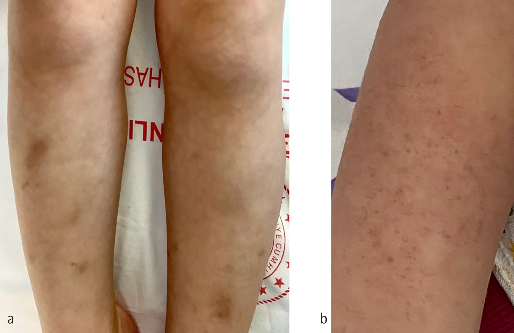
A 4-year-8-month-old girl was referred for evaluation of progressive gait disturbance and recurrent falls. She was born at term by cesarean section, weighing 3750 grams, with no perinatal complications. Her parents are first cousins.The initial symptoms emerged at 2.5 months of age as persistent diarrhea. By 1.5 years, she developed a livedo reticularis-like rash on the limbs, which became more pronounced in cold temperatures (), this was consistent with a mild acrocyanotic pattern previously described in the vascular manifestations of EE. Motor development was moderately delayed: she achieved head control at 2 months, sat at 12 months, and walked independently at 18 months. Speech developed between 18 and 24 months. She also had a known egg allergy. Family history was notable for neurodevelopmental delay in a male cousin. Neurological examination revealed hypotonia in the lower extremities and difficulty walking independently over short distances. Orthopedic evaluation demonstrated bilateral flat feet. Brain magnetic resonance imaging (MRI) showed a small T2/FLAIR hyperintense lesion in the left basal ganglia, consistent with a gliotic focus. Electromyography results were normal. Echocardiography revealed a small secundum atrial septal defect.
Biochemical analyses demonstrated consistently elevated serum lactate (3.2 mmol/L; reference <2.2) and markedly increased urinary ethylmalonic acid (up to 554.98 mmol/mol creatinine; reference <17). The acylcarnitine profile revealed a mildly elevated butyryl-carnitine (C4) level of 0.9192 µmol/L (reference range: 0-0.75 µmol/L). Molecular genetic testing identified a homozygous ETHE1 c.3G>T (p.Met1Ile) mutation, with both parents found to be heterozygous carriers. Based on these biochemical and genetic findings, the diagnosis of EE was established at the age of 3 years.
Following confirmation of the diagnosis, treatment was initiated with oral metronidazole (30 mg/kg/day) to reduce hydrogen sulfide production, N-acetylcysteine (30 mg/kg/day) to support detoxification pathways, riboflavin (10 mg/kg/day) and coenzyme Q10 (10 mg/kg/day) for mitochondrial support, and L-carnitine (50 mg/kg/day) to enhance energy metabolism. She also received biotin, multivitamin, zinc sulfate, and vitamin D supplementation. Within two months of treatment, she regained the ability to stand with support and crawl, having been previously unable to stand independently. However, by the fourth month, she lost this ability again, while petechiae and vascular skin changes not only persisted but also became more prominent during follow-up (). These cutaneous findings were characterized by non-blanching pinpoint purpura on the forearm, consistent with recurrent microvascular involvement. Differential diagnoses such as vasculitis or connective tissue disease, were considered but excluded based on the chronic course, lack of systemic inflammatory markers, and the presence of biochemical abnormalities specific to EE. The clinical course, developmental milestones, laboratory investigations, and imaging results are summarized in .
Table 1. Chronological summary of clinical, developmental, biochemical, and imaging findings in the patient
Age/date | Clinical findings | Developmental milestones | Laboratory results | Imaging/other investigations |
|---|---|---|---|---|
2.5 months | Persistent diarrhea | Head control at 2 months | — | — |
12 months | - | Sitting achieved | — | — |
18 months | Livedo reticularis-like rash on limbs, more pronounced in cold (mild acrocyanotic pattern) | Independent walking achieved | — | — |
18-24 months | - | Speech development | — | — |
3 y (at diagnosis, treatment started) | Progressive gait disturbance, recurrent falls, petechiae, vascular skin changes | Unable to walk independently (only a few assisted steps) | Serum lactate ↑; Urinary EMA 554.98 mmol/mol Crea (ref <17); Plasma C4 0.9192 µmol/L (ref 0-0.75) | MRI: small T2/FLAIR lesion (basal ganglia); EMG: normal; ECHO: small ASD |
3 y 7 m | Good general condition, petechiae + livedo rash, soft systolic murmur | Walking 15-20 m independently | Urinary EMA 171.41 mmol/mol Crea ↑; Plasma C4 0.81 µmol/L | — |
3 y 10 m | Good general condition with petechiae and livedo rash; mild reduction in walking distance | Independent walking only 8-10 steps | Urinary EMA 186.63 mmol/mol Crea ↑; Plasma C4 0.44 µmol/L | — |
4 y 3 m | Petechiae and livedo rash; pedal edema; facial telangiectasia | Lost ability to walk independently | Urinary EMA 237.41 mmol/mol Crea ↑; Plasma C4 0.98 µmol/L ↑ | — |
4 y 8 m | Persistent diarrhea (2-3 times/day); abdominal pain; petechiae and livedo rash; lower-limb spasticity and hyperreflexia | Stopped walking, only crawling | EMA 85.10 mmol/mol Crea ↑; C4 1.0365 µmol/L ↑ | — |
Treatment initiated at age 3 years. Serum lactate <2.2 mmol/L; urinary EMA <17 mmol/mol creatinine; plasma butyryl-carnitine (C4) 0-0.75 µmol/L. ASD: Atrial septal defect, ECHO: Echocardiography, EMA: Ethylmalonic acid, EMG: Electromyography, MRI: Magnetic resonance imaging
DISCUSSION
EE is classically described as a severe early-onset disorder characterized by rapidly progressive neurodegeneration, chronic diarrhea, petechiae, and acrocyanosis, with most patients succumbing within the first decade of life. However, accumulating evidence, including recent case reports and cohort analyses, suggests that EE can exhibit a broader clinical spectrum than previously appreciated. The patient described in this report exemplifies an atypically mild phenotype. Despite carrying a homozygous c.3G>T (p.Met1Ile) ETHE1 mutation predicted to result in complete loss of enzyme function, she demonstrated a protracted clinical course with no acute encephalopathic crises by nearly the age of five years.
This phenotypic heterogeneity has been well documented in recent literature. Kashima et al., described a child harboring a homozygous ETHE1 c.586G>A variant who exhibited only mild motor and speech delay, subtle biochemical abnormalities, and a normal MRI, reinforcing the notion that even pathogenic ETHE1 variants may yield attenuated clinical courses. Similarly, Ersoy et al., described an 11-year-old boy with the same c.3G>T variant as our patient who did not exhibit cognitive impairment or encephalopathic episodes but suffered from spastic paraparesis and chronic diarrhea. These cases, along with our patient, support the influence of modifier genes or residual enzyme activity in shaping disease severity. Environmental triggers, infection burden, and possible nuclear genetic modifiers have been hypothesized as additional contributors to this clinical variability, although definitive data are lacking.
The clinical progression in our patient parallels that of classical EE, though in a milder and slower course. Diarrhea emerged as the first symptom in early infancy, followed by vascular skin changes around 18 months and progressive gait disturbance in later childhood. This sequence aligns with the pattern proposed in classical EE, albeit in a slower and less aggressive form. Kasapkara et al. have emphasized the diagnostic significance of early gastrointestinal and cutaneous features, for instance, persistent diarrhea and livedo as early signs leading to EE diagnosis in two affected siblings. In our case, the diagnosis was delayed despite the presence of persistent diarrhea and vascular rash in infancy. This delay highlights the lack of awareness about the disease and the non-specific nature of early symptoms, which can be mistaken for common pediatric conditions. Increased recognition of these signs among pediatricians and dermatologists could reduce diagnostic delays.
Cutaneous manifestations, including livedo reticularis-like rash and recurrent petechiae, are frequent but often underrecognized features of EE. These can mimic vasculitis, collagen vascular disorders, or infectious purpura, leading to potential diagnostic delays. In our patient, the chronicity of lesions since infancy, their cold-induced exacerbation, and their coexistence with metabolic hallmarks (ethylmalonic aciduria, elevated C4) supported their attribution to EE. Therefore, careful dermatologic evaluation may provide an early diagnostic clue in atypical or milder phenotypes.
Biochemically, our patient displayed markedly elevated urinary ethylmalonic acid and mildly increased plasma C4 acylcarnitine, consistent with the metabolic fingerprint of EE. Hydrogen sulfide accumulation, resulting from deficient activity of the ETHE1-encoded mitochondrial sulfur dioxygenase, leads to secondary inhibition of short-chain acyl-CoA dehydrogenase and cytochrome c oxidase. This pathophysiological mechanism accounts for both the characteristic biochemical abnormalities and the multisystem clinical features observed in EE. The critical role of the ETHE1-encoded mitochondrial sulfur dioxygenase in mitochondrial hydrogen sulfide detoxification has been well established in previous studies.,
Treatment in EE remains primarily supportive, symptomatic. Our patient received continuous therapy for about two years with metronidazole, N-acetylcysteine, and mitochondrial cofactors such as riboflavin, coenzyme Q10, carnitine, and biotin. These agents aim to reduce hydrogen sulfide accumulation and support oxidative metabolism. Although not curative, they may slow progression and improve quality of life. In our case, transient motor improvements were noted after therapy initiation, consistent with previous reports describing partial clinical responses to metronidazole and N-acetylcysteine therapy., However, these improvements were reported by parents and clinical observation rather than standardized scales. The persistence of petechiae and gradual motor regression over time suggest that treatment may have stabilized but did not reverse disease activity. Notably, high-dose intravenous N-acetylcysteine has been reported to improve encephalopathic crises in EE patients, further supporting the therapeutic potential of such metabolic interventions, even in severe presentations.
The literature review by Platt et al. provides additional support for the existence of milder phenotypes. In their cohort of 70 patients, eight cases were classified as mild, characterized by slower neurological progression and lower urinary ethylmalonic acid and C4 acylcarnitine levels compared with classical cases. Among the reported genotypes, the ETHE1 c.3G>T (p.Met1Ile) variant was one of the most frequent and was exclusively associated with mild presentations, as no patients harboring this variant exhibited a classical or severe phenotype. Importantly, the current age of patients with mild phenotypes was significantly higher, indicating better long-term outcomes. The study also emphasized that symptom onset in milder cases is often dominated by gastrointestinal and vascular manifestations rather than acute neuroregression, further supporting our observations.
In summary, this case contributes to the literature on EE in several important ways. First, it reinforces the genotype-phenotype correlation by demonstrating that the recurrent ETHE1 c.3G>T (p.Met1Ile) mutation, typically associated with severe disease, can manifest with a mild phenotype. Second, it highlights the diagnostic value of early gastrointestinal and vascular manifestations, which in our patient preceded neurological decline and could help shorten diagnostic delays if recognized. Recognizing these early findings may enable earlier metabolic intervention and prevent irreversible neurological damage. Third, it underscores the risk of misattributing these early non-neurological signs to common pediatric conditions, emphasizing the need for awareness among pediatricians and dermatologists. Finally, it provides additional evidence that continuous administration of metronidazole, N-acetylcysteine, and mitochondrial cofactors may help stabilize the clinical course, even if they do not reverse disease activity. Taken together, these observations strengthen our understanding of the phenotypic spectrum of EE and offer practical insights for diagnosis and management.
(a) Livedo reticularis-like discoloration on the anterior aspects of both lower limbs, more pronounced in cold temperatures, consistent with a mild acrocyanotic pattern previously described in EE. Differential diagnoses such as vasculitis, connective tissue disorders, and infectious etiologies were considered. However, the persistence of the rash since infancy, absence of systemic inflammatory markers, and concurrent metabolic abnormalities supported attributing it to EE-related microangiopathy.
(b) Petechial lesions on the right forearm observed four months after treatment initiation (age 3 years 4 months). Multiple non-blanching, pinpoint purpuric macules are visible, consistent with recurrent cutaneous microvascular involvement. These findings coincided with loss of previously regained motor abilities, while petechiae and vascular skin changes persisted and became more prominent during follow-up, illustrating the chronic and fluctuating course of the disease.
Footnotes
Ethics
Informed Consent: Informed consent was obtained from the patients’ family and/or patients in the study.
Authorship Contributions
Surgical and Medical Practices: M.Y.Ç., T.Ç., Concept: M.Y.Ç., Design: M.Y.Ç., B.K., E.B., Data Collection or Processing: M.Y.Ç., B.K., E.B., T.Ç., Analysis or Interpretation: M.Y.Ç., Literature Search: M.Y.Ç., Writing: M.Y.Ç.
Conflict of Interest: The authors have no conflicts of interest to declare.
Financial Disclosure: The authors declared that this study received no financial support.
References
Tiranti V, Diodato D, Lamantea E, Accarino G, Fernandez-Vizarra E, Mora M, Zeviani M. ETHE1 mutations are specific to ethylmalonic encephalopathy. J Med Genet. 2006;43(4):340-346.
Govindaraj P, Vedantham S, Chacko A, Vasudevan A. Child neurology: ethylmalonic encephalopathy. Neurology. 2020;94(12):e1336-e1339.
Ersoy M, Tiranti V, Zeviani M. Ethylmalonic encephalopathy: clinical course and therapy response in an uncommon mild case with a severe ETHE1 mutation. Mol Genet Metab Rep. 2020;25:100641.
Kashima DT, Sloan-Heggen CM, Gottlieb-Smith RJ, Barone Pritchard A. An atypically mild case of ethylmalonic encephalopathy with pathogenic ETHE1 variant. Am J Med Genet A. 2023;191(6):1614-1618.
DiMauro S, Schon EA. Mitochondrial respiratory-chain diseases. N Engl J Med. 2003;348(26):2656-2668.
Chen C, Chen Y, Guan M-X. A peep into mitochondrial disorder: multifaceted from mitochondrial DNA mutations to nuclear gene modulation. Protein Cell. 2015;6(12):862-870.
Platt I, Bisgin A, Kilavuz S. Ethylmalonic encephalopathy: a literature review and two new cases of mild phenotype. Neurol Sci. 2023;44(11):3827-3852.
Yiş U, Hız Kurul S, Duman O, Yılmaz C, Dirik E. Importance of acrocyanosis in delayed walking. J Pediatr Neurosci. 2015;10(1):80-81.
Kasapkara ÇS, Güzel-Ozantürk A, Dursun A, Sivri HS, Tokatlı A, Coşkun T. Siblings with ethylmalonic encephalopathy: case report. Turk J Pediatr. 2018;60(1):104-107.
Zafeiriou DI, Augoustidou-Savvopoulou P, Haas D, Lehnert W, Vargiami E, Tsantali C, Sewell AC. Ethylmalonic encephalopathy: clinical and biochemical observations. Neuropediatrics. 2007;38(2):78-82.
Luna-Sánchez M, Díaz-Casado ME, Barca E, Tejada MA, Montilla-García Á, Cobos EJ, Escames G, Acuña-Castroviejo D, Quinzii CM. CoQ deficiency causes disruption of mitochondrial sulfide oxidation, a new pathomechanism associated with this syndrome. EMBO Mol Med. 2017;9(1):78-95.
Viscomi C, Burlina AB, Dweikat I, Savoiardo M, Lamperti C, Hildebrandt T, Tiranti V, Zeviani M. Combined treatment with oral metronidazole and N-acetylcysteine is effective in ethylmalonic encephalopathy. Nat Med. 2010;16(8):869-871.
Kılıç M, Tokatlı A, Dursun A, Coşkun T, Sivri HS. Successful treatment of a patient with ethylmalonic encephalopathy by intravenous N-acetylcysteine. Metab Brain Dis. 2017;32(2):293-296.